Nasolacrimal Duct Obstruction (NLDO)

Where do tears come from?
Under normal circumstances the eye is kept moist by tears produced by glands on the surface of the eye. When something irritates the eye, or when we get upset enough, the lacrimal gland (located in the eye socket, above the eyeball) releases a burst of water and we cry.

Where do tears go?
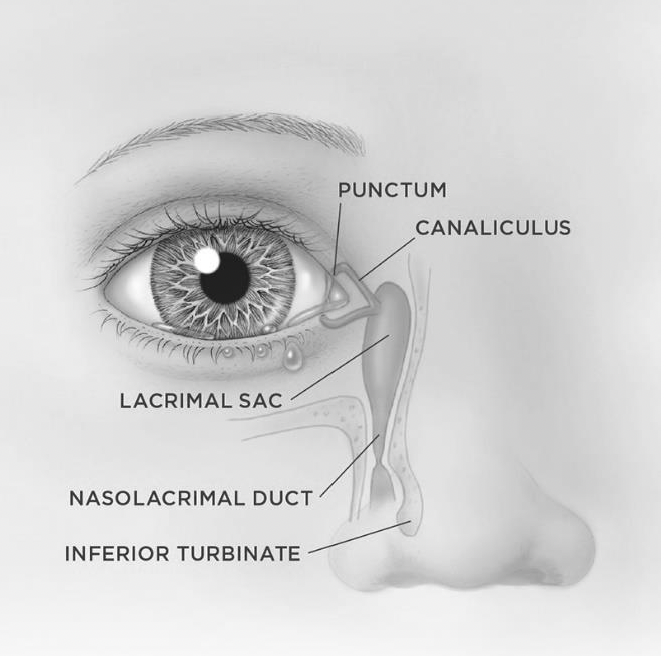
Along each eyelid, near the nose, is a tiny hole (punctum) – the punctum is the entry point to the tear drainage system. Each punctum is connected to a tiny tube that runs within the eyelid to the nose (canaliculus). The canaliculus connects to the lacrimal sac, the spongy structure at the “corner” of the eye by the nose.
Finally, the lacrimal sac leads to the tear duct, which runs deep within the bone and empties into the nose.

What causes a blocked tear duct in children?

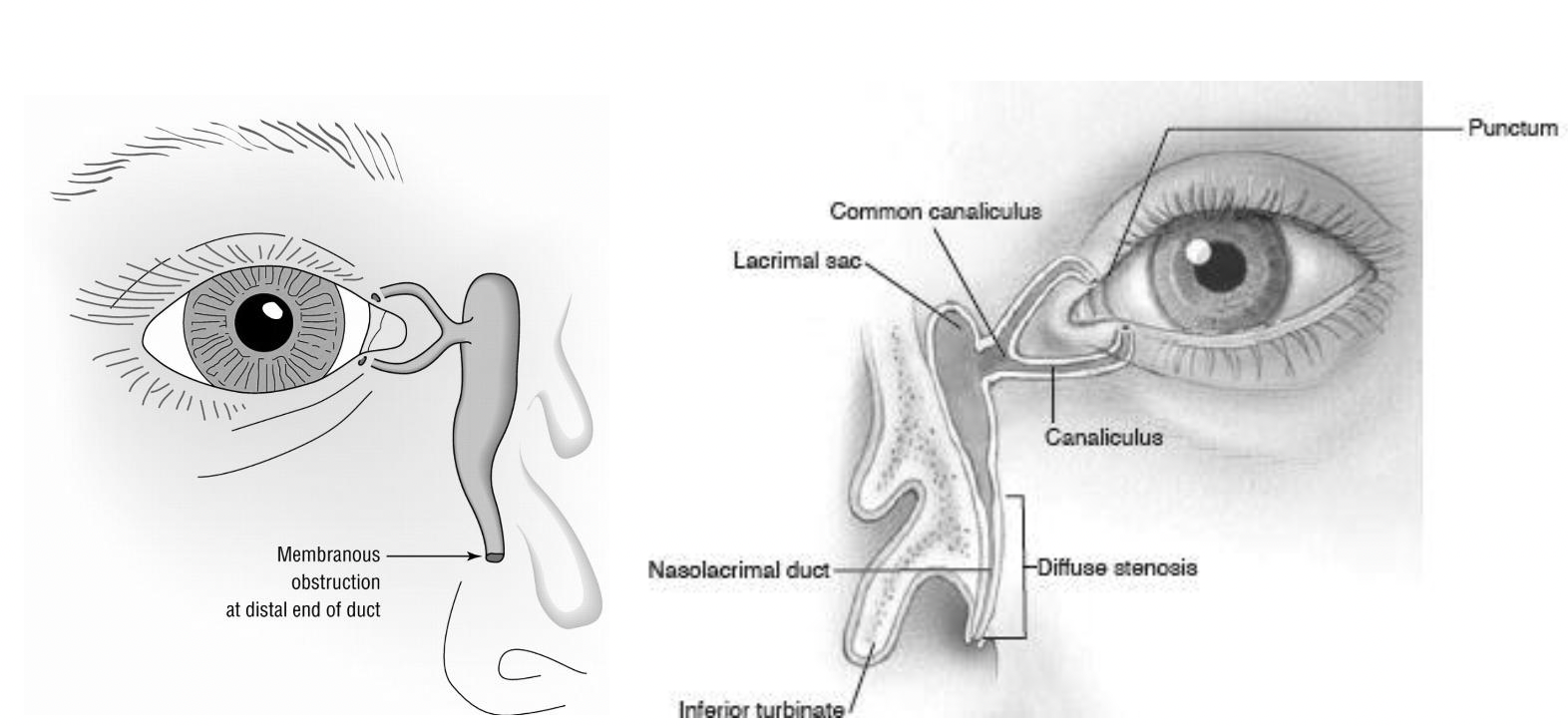
During fetal development, the tear duct begins as a solid cylinder of cells within the facial bones. These tear duct cells communicate with each other to form a hollow tube within the bone: the nasolacrimal duct. Sometimes, this duct is not completely open at birth – most often, this is because of a thin membrane running across the duct that prevents the tears from freely flowing (imagine a drumhead). Sometimes, the bone surrounding the duct is tight (stenosis), pinching off the flow of tears. Approximately 5% of newborns have a NLDO.

When the tear duct does not properly empty into the nose, the tears back up and overflow (epiphora). Another thing: the tear duct lining is like the lining of the nose – it produces mucous. If the tear duct drainage is abnormal, this mucous backs up instead of going into the nose.

Therefore, there can be a yellow-white discharge on the lashes and around the eye. This discharge poses no threat to visual development or to the eye tissues although it can be irritating to the skin and is not very attractive.

What To Do?

Of the 5% of newborns born with a NLDO, around 90% get better on their own by the age of 12 months– the membrane occluding the duct opens spontaneously or the bone grows and the duct is no longer choked off. (For every 1,000 babies born, 50 have NLDO; by age 12 months, 5 of these 50 will still have an obstruction). The odds are heavily in favor of spontaneous resolution.

Until an NLDO spontaneously resolves, there are measures you can take to minimize the build-up of the “gooey” discharge. First and foremost, gentle massage of the lacrimal sac can be performed. This creates pushes out the mucous and debris that have built up within the obstructed tear duct system. To massage the lacrimal sac, gently press then release, the little bump at the corner of the eyelids where it meets the nose (this round spongy structures give a little and the baby will not feel any pain. Performing this massage before each diaper change will usually keep the symptoms under control. This will NOT cure the obstruction, but it will reduce the gooey discharge while you wait for the baby’s tear duct to enlarge over time.

Sometimes there is bacterial overgrowth within the blocked tear duct. Then (and only then) antibiotics can be used. As you may know, the inappropriate and indiscriminate use of antibiotics has led to the bacteria that can no longer be easily killed by antibiotics. If you need to use antibiotic eye drops, then please take care to use them as directed. For NLDO with secondary infection use one drop 2 times a day for 5 days, then try to go back to massage alone.

Finally, there are surgical options to open the obstruction. Inserting a metal probe into the nasolacrimal duct will open any membranes and allow the surgeon to feel whether or not the bone around the duct is too tight. This can be done safely in the office if the baby is small enough to be held relatively still; the ideal age is 6 to 9 months. The benefits of in-office NLDO probing include an excellent chance of immediate cure (95%) and the avoidance of a possible general anesthesia (see below: “What if the NLDO doesn’t go away on it’s own by the first birthday?”). Other benefits include money and time saved (fewer eye drops, less time massaging, and no need to take off from work for a trip to the operating room). The alternative is to wait for spontaneous resolution. If there is no spontaneous resolution by 13-14 months, the NLDO is unlikely to resolve on its own and a trip to the operating room is the only way to fix it. The risks of in-office probing include failure to achieve cure, infection, nosebleed, discomfort, and damage to the nasolacrimal drainage system; these same risks apply when the procedure is performed in an operating room. If, at the time of in-office probing, it becomes unsafe to proceed (baby moves too much, etc.) then the procedure is immediately stopped. Probing the nasolacrimal duct on a person who is awake is not painful, but it is by no means a pleasurable sensation. Adults with tear duct problems are routinely probed in the office and most describe it feeling like soda bubbles going up the nose.

How is a nasolacrimal duct probed?

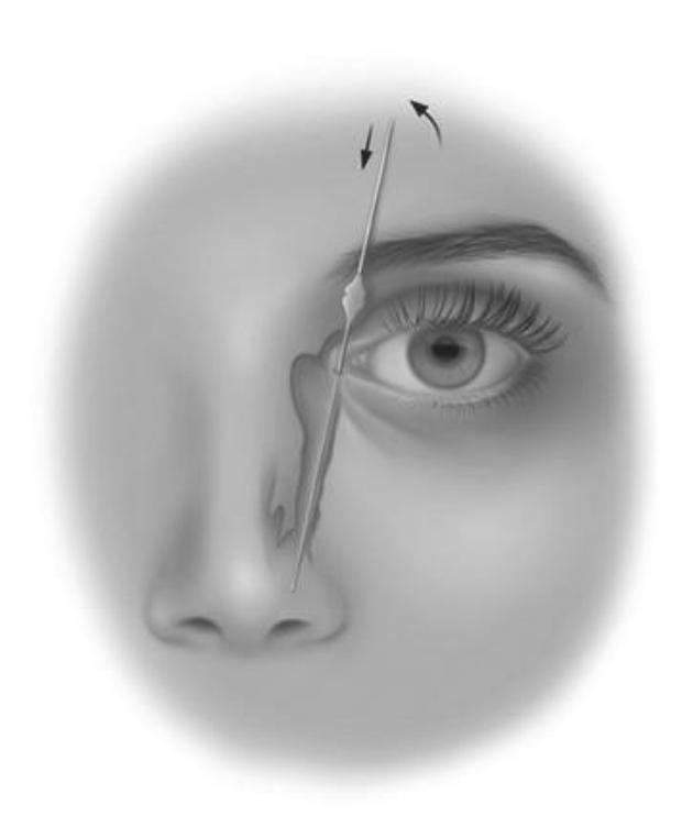
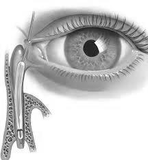
This drawing demonstrates how the nasolacrimal duct is probed. First, the probe (a wire with a rounded tip) is placed into the punctum and canaliculus. Then, the probe is directed down into the duct. As you can see here, the probe exits underneath one of the nose bones.

Notice that the nasolacrimal duct exits underneath one of the nose bones (inferior turbinate). If the turbinate is pressing against the duct, then tears cannot flow. On occasion, it is necessary to fracture the bone out of the way (not remove it). When the bone is fractured, it does not alter the appearance of the nose in any way.

What if the NLDO doesn’t go away on its own by my baby’s first birthday?

If an NLDO persists beyond 13th or 14th month of age, then surgery in the operating room is an option (these children are too big for in-office probing). The cure rate from probing alone after 13 months is about 75%. One possible reason for this significant decrease in the success rate is that babies who do not get better on their own by 13 months are more likely to have something more than a simple membrane blocking the duct. These babies are more likely to have several membranes and/or bony stenosis. It is impossible to tell what is causing a NLDO just by looking at the patient; the surgeon must literally feel what the probe is doing to interpret the cause of the obstruction.

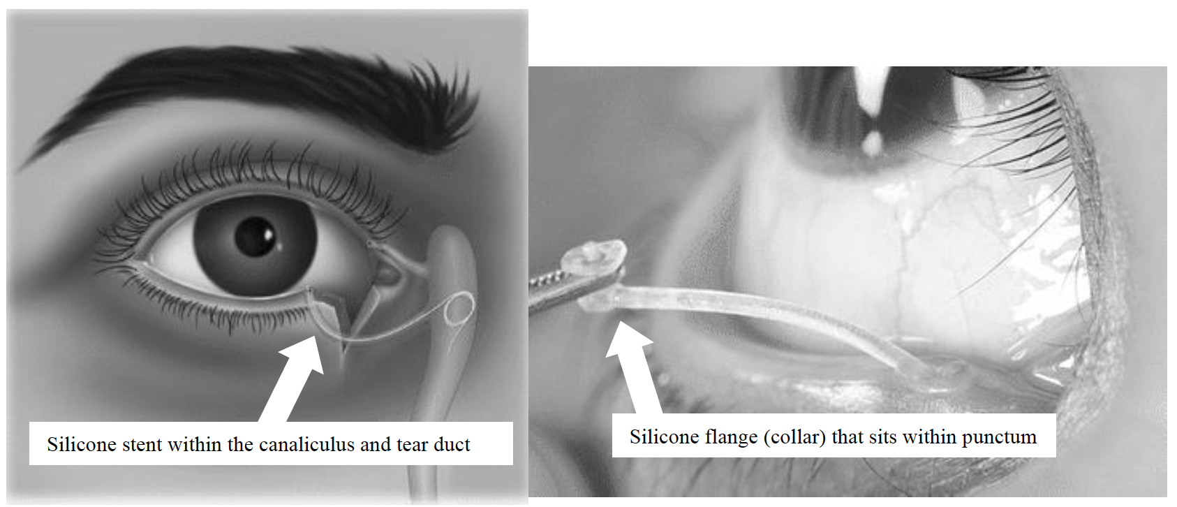
In the operating room a silicone tube (stent) is usually placed within the tear duct. This tube (ideally) stays in place for 6 months; we remove the tube in the office. The silicone tube sits flush within the opening in the eyelid (canaliculus) and the child feels nothing. The tube is completely external to the eye and is not sewn into anything.

The risk of general anesthesia is minimal for a healthy baby. The risk of nasolacrimal stent surgery includes failure to achieve cure, infection, nosebleed, discomfort, and damage to the nasolacrimal drainage system, but such complications are rare.

Balloon Dacryoplasty

If obstruction persists despite trying a stent and a repeat visit to the operating room is in order, the tear duct can be expanded using a balloon procedure (dacryoplasty). The balloon is placed through the punctum and expanded within the duct, then removed. This expands the diameter of the duct and the surrounding bone.

The risk surgery includes failure to achieve cure, infection, nosebleed, discomfort, and damage to the Nasolacrimal drainage system, but such complications are rare.

Enhance Your Child’s Vision and Confidence

If your child shows signs of NLDO, it’s essential to seek treatment early. Contact us today to schedule a consultation and discover how our specialized ptosis correction procedures can improve your child’s vision and appearance.