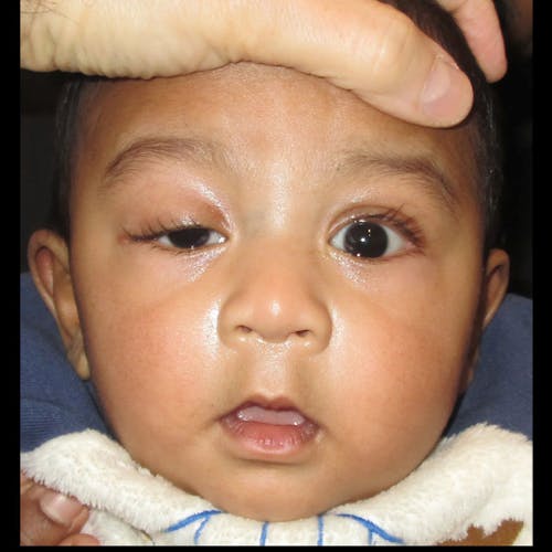
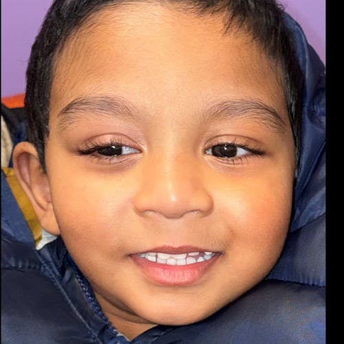
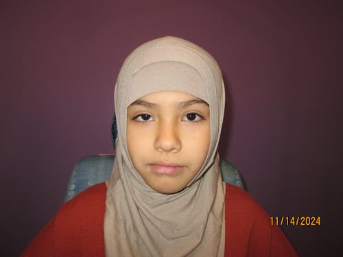
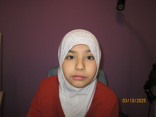
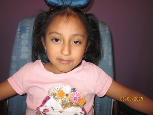
Advanced Ptosis Correction for Improved Vision and Appearance

A “droopy” or “lazy” eyelid is called ptosis. Older adults develop ptosis as part of the aging process because the tissues that hold up the eyelid stretch over time. Babies are born with ptosis because the muscle that lifts the lid did not develop properly. Pediatric ptosis can also have a neurologic basis. It is important to distinguish congenital (“myogenic”, muscular) ptosis from that caused by a neurological problem.

Apart from “looking different,” the medical risk of pediatric ptosis is permanent vision loss that can result when the lid covers the eye, preventing the brain from receiving information from that eye. Such vision loss is called amblyopia. While amblyopia can be reversed until 12 years of age, the underlying cause needs to be addressed. In other cases of ptosis, when the pupil is NOT completely covered, the ptosis can also cause significant astigmatism by pressing on the cornea. Finally, some babies with ptosis place their chin upwards (torticollis) as soon as they are capable of holding their own neck. This allows them to peek under the lid and use both eyes. When ptosis causes amblyopia, high astigmatism and/or a chin-up head position, timely surgery is indicated and any amblyopia present needs to be treated.

Congenital Ptosis
Congenital ptosis occurs because of abnormal development of the muscle and connective tissue that lift the eyelid. This can be associated with certain syndromes but usually is not. One of the hallmarks of congenital ptosis is the affected lid does not come all the way down when the eyes are looking down; the ptotic lid “lags” behind the normal lid (lid lag). Another characteristic is a relatively smooth lid with poor formation of the upper eyelid crease.

Neurologic Ptosis
All muscles in the body do as they are told by their nerve supply. Naturally, this applies to the muscles that rotate the eye (extraocular muscles) and those that lift the eyelid (levator and Muller muscles). If a neurologic problem affects the nerve supply of the levator – and ptosis results – this can be accompanied by abnormal function of the pupil and some of the extraocular muscles.

In addition to nerve impairment causing ptosis, there can be chemical abnormalities at the junction between the nerve and the muscle (myasthenia gravis). Myasthenia gravis is rare in childhood but needs to be considered.

Timing of surgery
Ptosis can cause amblyopia if the lid completely covers the pupil. In these cases, the ptosis must be repaired as soon as possible to prevent worsening of amblyopia and allow for its treatment. Another
indication for surgery is if the ptosis is causing high astigmatism (which can also produce amblyopia). Finally, if a child is lifting her chin all of the time she should have her ptosis repaired without significant delay. The abnormal head position (torticollis) is assumed so that she can use both eyes together and see things three-dimensionally.

If the ptosis is not severe enough to cause amblyopia there is nothing wrong with waiting until a child is 4 or 5 years old before surgery. Children younger than this don’t usually notice their appearance as different and are unlikely to be teased by other children. That said, some parents want earlier surgery and this is often a reasonable option.

Ptosis Surgery

There are two ways to correct ptosis. The first technique is to strengthen the levator or Muller muscle. Strengthening the levator usually involves an incision on the skin of the eyelid; for a Muller strengthening the incision is on the undersurface of the eyelid. There are benefits, risks and alternatives to each of these approaches, and each case needs to be determined individually.

The second technique is to mechanically lift the eyelid, bypassing the levator and Muller muscle function altogether. This involves placing material under the skin to suspend and hold the lid in place. There are several types of material used and all have a failure rate of about 20%. Synthetic materials are most commonly used but we also use tissue from the thigh (fascia lata) of a person who has died and made a gift. Fascia lata is tested for all known transmissible diseases and then irradiated. The risk of disease transmission is extremely low.

The greatest risk of ptosis surgery is the need for reoperation. Everybody heals differently. While some children require just one surgical repair – and this is what everyone involved would like – a fair number need more surgeries. It is impossible to predict who needs more than one surgery or the interval between surgeries. Basically, most children need just one repair but some need more than one.

There is a risk of infection, though this is not common. A very aggressive infection could cause vision loss or systemic infection but this is very rare. To minimize infection risk, oral antibiotics are used after frontalis suspension surgery, and all patients examined during the first week after surgery (when the risk of infection is greatest).

Just after surgery the eyelid is often higher than its final position. This is more typical with frontalis suspensions. The higher lid position can cause dryness of the eyeball when the patient is sleeping, so it is very important to keep the eye lubricated in the immediate post-operative period. As noted above, people with congenital ptosis have a problem with the lid coming down normally and you may have noticed that the eye with ptosis appears open when your child sleeps. All ptosis repair techniques have a tendency to exacerbate this inability to close the lid and will be noticeable when the patient is asleep or when she looks down; this is particularly the case when a suspension procedure is performed.

Enhance Your Child’s Vision and Confidence

If your child shows signs of ptosis, it’s important to seek treatment early. Contact us today to schedule a consultation and discover how our specialized ptosis correction procedures can improve your child’s vision and appearance.