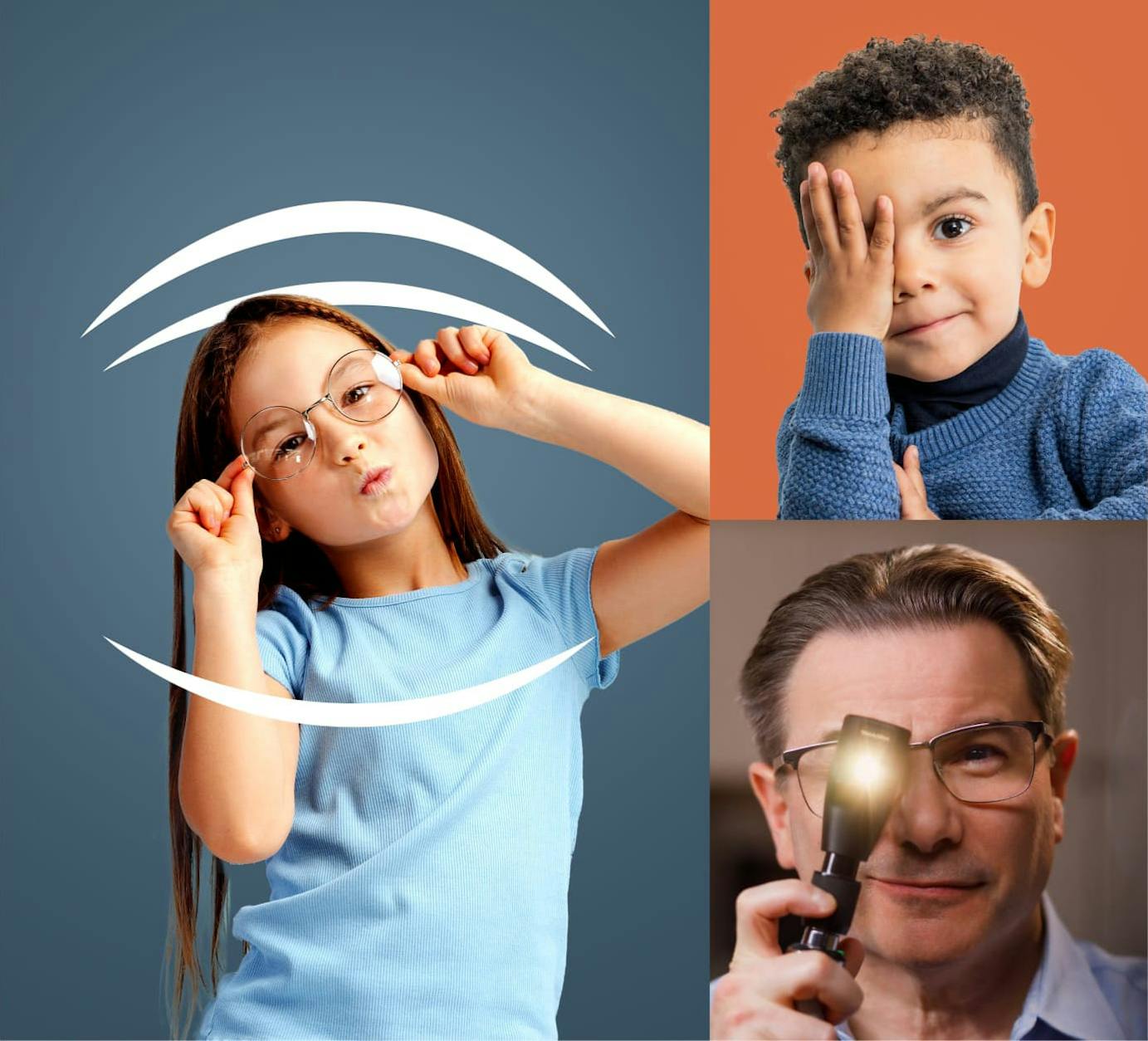
Vision is not an innate sense; it is acquired. While it may appear to “just happen” in most children, normal visual development depends on a precisely coordinated neurologic and optical process. Disruptions during this maturation period can lead to long-term visual consequences, underscoring the importance of early identification and appropriate management of refractive error in pediatric care.
The Physiology of Visual Development
Under normal conditions, the cornea and crystalline lens focus incoming light into a sharp image on the retina. The retina then converts this image into a neural signal, which is transmitted via the optic nerve to the occipital cortex, where visual perception occurs.
During the critical period of visual development—birth through approximately 8 years of age—consistent, accurate stimulation of the visual cortex is required to establish normal vision. When light is improperly focused onto the retina, resulting in a blurred image, the cortex receives degraded input.
A key concept during this period is interocular cortical competition:
- When one eye provides a clearer image than the other, the brain preferentially processes that input
- Cortical pathways corresponding to the blurred image are actively suppressed
- Over time, this suppression leads to the underdevelopment of the affected pathway
This process can result in amblyopia, characterized by:
- Reduced visual acuity
- Deficits not correctable with refractive lenses alone
Importantly, amblyopia is reversible if identified and treated early. Effective management during the critical period includes:
- Timely correction of refractive error
- Identification and treatment of amblyogenic conditions
- Restoration of balanced cortical stimulation between both eyes
When addressed appropriately, normal cortical development—and functional vision—can be achieved.
Types of Refractive Error
Understanding refractive error is fundamental to determining when intervention is necessary.
Myopia
In myopia, distant objects are focused in front of the retina rather than directly on it. The result is blurred distance vision, while near vision remains relatively intact.
Younger children often tolerate mild myopia (approximately –0.50 to –1.50 diopters) without functional impairment, as their visual demands are primarily near-based.
For this reason, low levels of myopia in early childhood may not require immediate correction unless they interfere with activities of daily living (ADLs) or contribute to amblyopia.
Hyperopia
Hyperopia occurs when light is focused behind the retina. In children, this is often physiologic. The accommodative ability of the crystalline lens allows the eye to “pull” the image forward onto the retina.
The typical physiologic range of hyperopia in children is between plano (0.00 D) and +4.00 D. Beyond this range, however, the risk of accommodative esotropia and amblyopia increases.
Clinical decision-making must account for both the magnitude of hyperopia and the child’s ability to compensate.
Astigmatism
Astigmatism results from an irregular curvature of the cornea or lens, causing light to focus at multiple points rather than a single focal point.
Depending on the configuration, one focal point may lie on the retina while another lies in front of or behind it, or both may be off the retinal plane.
Clinically significant astigmatism can degrade image quality and, if uncorrected, contribute to amblyopia during the critical period of development.
The directional axis of the astigmatism:
| Physiologic range: | Examples of Clinical Notation: |
|---|---|
| Myopia: none. | - 4.00 D sphere |
| Plano (no refractive error) | Plano |
| Hyperopia: 0.00 D to + 4.00 D | +3.25 D sphere |
| Astigmatism: 0.00 D to + 2.00 D | “pure” astigmatism: plano +3.00 x 90 Myopic astigmatism: -3.50 + 1.75 x100 Hyperopic astigmatism: +5.75 +2.00 x80 |
* D = diopter (unit of measurement for refraction)
Anisometropia and Amblyopia Risk
Anisometropia refers to a difference in refractive error between the two eyes. Even when each eye falls within the physiologic range, a significant interocular discrepancy can lead to cortical suppression of the more ametropic eye.
Hyperopic anisometropia is particularly amblyogenic. For example, a child with +2.00 D in one eye and plano in the other may preferentially use the clearer eye, leading to suppression and eventual amblyopia in the hyperopic eye.
Clinical thresholds for concern include:
- ≥3.00 D difference in myopia
- ≥1.50 D difference in hyperopia
- ≥2.00 D difference in astigmatism
Determining Refractive Error in Pre-Verbal Patients
Objective measurement of refractive error in infants and young children is performed using cycloplegic retinoscopy. Cycloplegic agents (topical cholinergic antagonists) temporarily paralyze accommodation and dilate the pupil, allowing accurate assessment of the eye’s refractive state.
Approximately 30 minutes after they are instilled, a retinoscope is used to project a streak of light onto the retina. The reflected light is analyzed to determine refractive error. While automated infrared-based systems are available, they may involve trade-offs in accuracy and are not universally preferred.
Guidelines for Prescribing Eyeglasses in Infants and Pre-School Children
There is no universal “cookbook” for prescribing glasses in young children. Clinical judgment must integrate cycloplegic findings, visual acuity (when measurable), presence of amblyopia, and functional impact on ADLs.
In general, if visual acuity is equal between both eyes and there is minimal anisometropia, glasses may not be necessary for:
- Myopia less than –2.00 D
- Hyperopia less than +4.50 D
- Astigmatism less than +2.50 D
However, even in the absence of measurable acuity differences, glasses may be indicated in the following scenarios:
- Hyperopia ≥ +4.50 D (even without esotropia)
- Anisometropia exceeding the thresholds noted above
If amblyopia is present and visual acuity cannot be equalized with a trial lens, any significant refractive error must be corrected.
Guidelines for Children Able to Participate in Visual Acuity Testing
Once a child can reliably participate in visual acuity testing, subjective input becomes a useful—but not exclusive—component of clinical decision-making.
General benchmarks include:
- Age 4: 20/40 or better in each eye (tested monocularly).
- Age 6: 20/30 or better.
- Age 8: 20/20 expected.
If a child meets these thresholds without correction and has no significant refractive error or anisometropia, glasses are typically not indicated.
However, visual acuity alone should not dictate management. Cycloplegic findings, binocular function, and risk of amblyopia remain critical considerations.
When to Refer for Ophthalmologic Evaluation
Early detection remains the most effective strategy for preventing permanent vision loss.
All children should undergo formal visual acuity screening by their fourth birthday. Despite this, only a minority of children in the United States are screened within this timeframe.
Referral to a pediatric ophthalmologist is indicated when:
- Visual acuity is worse than 20/40 in one or both eyes
- There is a difference of two or more lines between eyes
- Clinical findings such as strabismus, nystagmus, or an abnormal red reflex are present
Notably, screening does not require specialized equipment or subspecialty training. A calibrated picture chart and a few minutes are sufficient to identify most at-risk children.
Screening Recommendations
Joint guidelines from the American Academy of Ophthalmology and the American Association for Pediatric Ophthalmology and Strabismus emphasize early and repeated screening:
- Newborns should be examined for ocular health, with referral for high-risk infants
- All infants should undergo screening by 6 months of age
- Vision screening should occur around age 3½, with emphasis on acuity
- At age 5, both visual acuity and ocular alignment should be assessed
- Additional screening should occur during school years or if symptoms arise
Most serious, treatable pediatric eye conditions are identifiable during the preschool years. Many are associated with family history, underscoring the importance of vigilance in at-risk populations.
In Conclusion
Prescribing eyeglasses in pediatric patients requires more than interpreting refractive error. It demands an understanding of visual development, cortical plasticity, and the dynamic interplay between the two eyes during the critical period.
For pediatricians, early screening and timely referral are the most impactful interventions. By identifying children at risk and facilitating prompt ophthalmologic evaluation, it is possible to prevent amblyopia and ensure normal visual development.