
View Other Patients
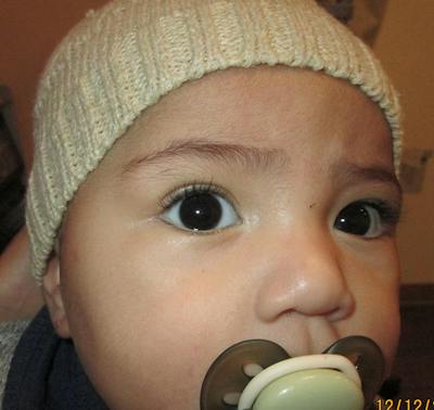
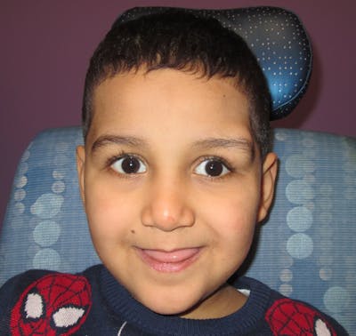
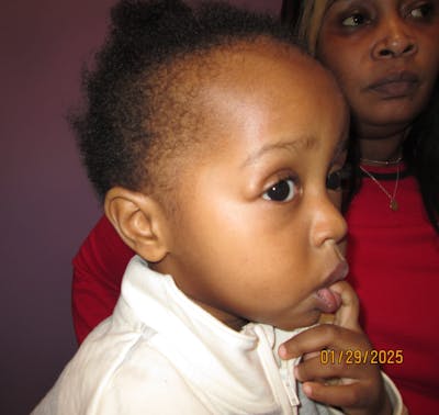
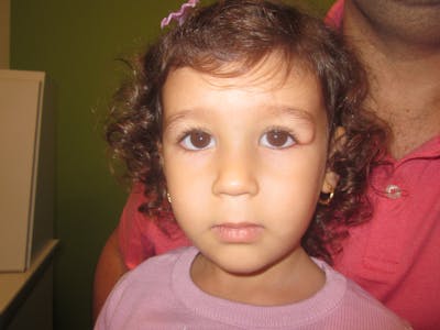
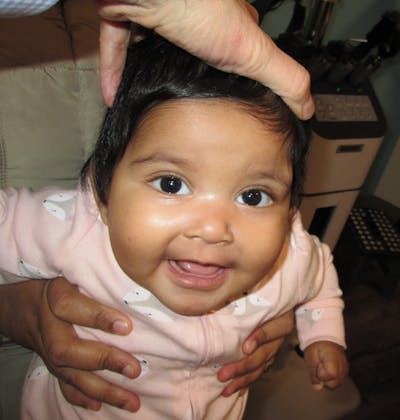
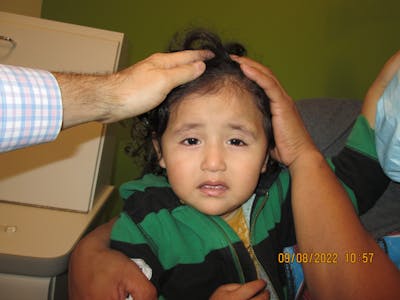
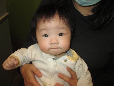


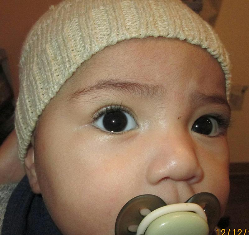
Before Surgery
Jenny presented with a cyst that had been slowly growing in her upper eyelid. The vast majority of childhood eyelid and orbital masses are benign. Common examples include chalazion, pilomatricoma, and dermoid cysts. Based on Jenny's history and examination, this lesion was most consistent with a dermoid cyst.
Dermoid cysts are benign developmental lesions that form in utero when a tiny portion of skin becomes trapped beneath the surface during early development. Although present from birth, they often become more noticeable as a child grows.
In this case, the cyst created a visible fullness of the upper eyelid. These lesions are typically harmless, but they usually enlarge slowly over time and do not resolve on their own. Aside from cosmetic concerns, one of the main risks is rupture following trauma. If a dermoid cyst ruptures, its contents can spill into the surrounding tissues and trigger significant inflammation, making subsequent surgery more difficult and potentially increasing the risk of irregular scarring.
The goal of surgery is to remove the cyst completely while preserving the normal appearance and function of the eyelid and surrounding tissues.
After Surgery
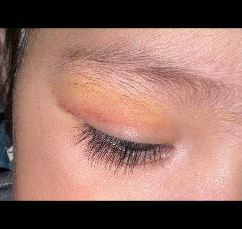
The dermoid cyst was successfully removed through the eyelid crease incision. As healing progresses, the incision typically becomes increasingly difficult to see because it lies within a natural skin crease.
The post-operative photograph demonstrates restoration of the normal contour of the upper eyelid and brow region, with a natural appearance and minimal visible evidence of surgery. Most children recover quickly and return to normal activities the same day.
Recovery & Results
Every child heals differently, and individual results will vary. Temporary swelling, bruising, redness, and mild eyelid asymmetry are common during the early healing period and generally improve with time.
The photographs shown represent one child's surgical journey and are provided for educational purposes. They should not be viewed as a guarantee of a specific outcome for any other patient.
During consultation, I discuss the expected recovery process, potential risks, and anticipated results for each child based on their individual anatomy and condition.